Electrophysiology on brain slices

Managers: Clémentine Bosch-Bouju, Xavier Fioramonti, David Jarriault

Applications

  • Measurement of synaptic transmission and plasticity
  • Measurement of electrophysiological parameters and neuronal excitability on rodent brain slices
  • Measurement of neuron sensitivity to nutrients and hormones
  • Specific studies of particular neurons using “fluorescent mice” (NPY-GFP, POMC-GFP, D1R-GFP, D2R-GFP, Pet1-mCherry, CxCR1-GFP, etc.).
  • Brain structures studied: hippocampus, cortex, nucleus accumbens, striatum, dorsal raphe, hypothalamic nuclei
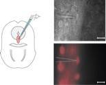
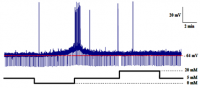
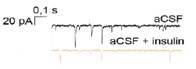
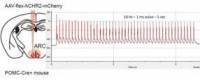
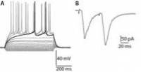
Infrared (top) and fluorescence (bottom) imaging of serotonin (5-HT) neurons in the dorsal raphe of Pet1-cre/mCherry mice
Current-clamp electrical recording of a NPY-GFP neuron in response to variations in glucose concentration.
Voltage-clamp electrical recording of spontaneous GABA inhibitory currents on dorsal raphe 5-HT neurons in response to insulin
Optogenetic stimulation of POMC neurons in the arcuate nucleus of POMC-cre mice injected with AAV-flex-hCHR2-mCherry
Electrophysiological recording of a striatal middle spiny neuron in mice. A, Electrophysiological profile of the neuron following injection of current jumps (10 pA increment). B, Synaptic response of the neuron following stimulation of glutamatergic afferents (2 stimulations at 50 ms intervals).

Equipment

  • 3 brain slice patch-clamp electrophysiology stations
  • Upright microscopes with white light, infrared and fluorescence (combinable LEDs 370 nm, 460nm, 580nm)
  • Electrical stimulation
  • Optogenetic stimulation
Photos of the NutriEphy electrophysiology room